Histology of Epithelium, Connective Tissue, Muscle, Cartilage, Glands, GIT, Liver and Respiratory Tract
Below is a **single-place, SEO-optimized, exam-ready histology guide** covering **epithelium, glands, connective tissue, fibers, cartilage, muscle, lymphatic system, GIT, liver, and respiratory tract**.
Structured with **clear H1–H3 headings**, **high-yield features**, and **clinical correlations** for medical students and competitive exams.
---
# Histology of Human Tissues and Organ Systems – Complete Guide
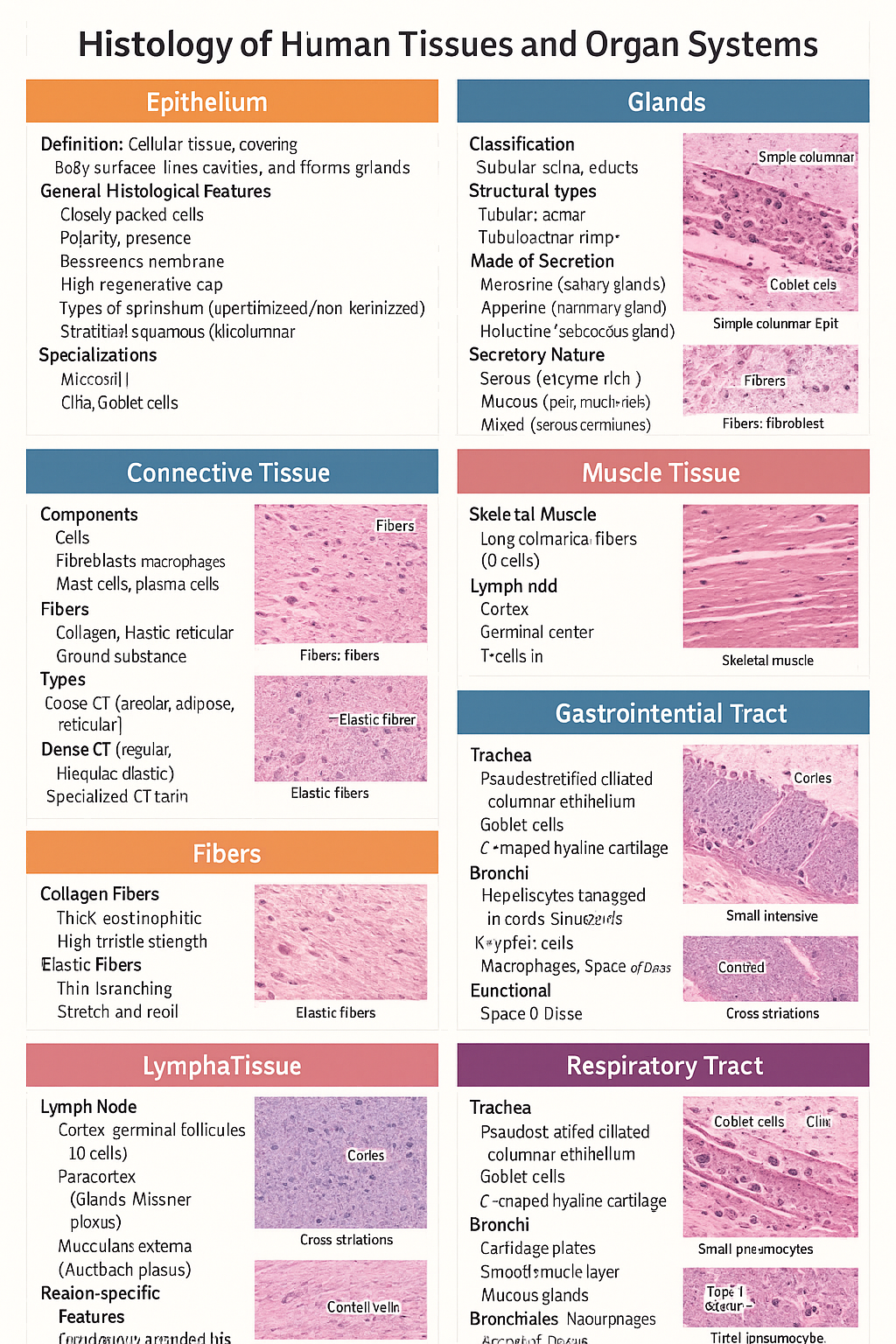
## Histology of Epithelium
### Definition
Epithelium is a **cellular tissue** that covers body surfaces, lines cavities, and forms glands.
### General Histological Features
* Closely packed cells with **minimal extracellular matrix**
* **Polarity**: apical, lateral, basal surfaces
* **Basement membrane** present
* **Avascular**, richly innervated
* High **regenerative capacity**
### Types of Epithelium
* **Simple squamous**: alveoli, capillaries
* **Simple cuboidal**: kidney tubules, glands
* **Simple columnar**: stomach, intestine
* **Stratified squamous (keratinized/non-keratinized)**: skin / oral cavity
* **Pseudostratified ciliated columnar**: trachea
* **Transitional epithelium**: urinary bladder
### Specializations
* **Microvilli** – absorption
* **Cilia** – movement
* **Goblet cells** – mucus secretion
---
## Histology of Glands
### Classification
* **Exocrine glands** – ducts present
* **Endocrine glands** – ductless, secrete into blood
### Structural Types
* **Tubular**, **acinar**, **tubuloacinar**
* **Simple** or **compound**
### Mode of Secretion
* **Merocrine** (salivary glands)
* **Apocrine** (mammary gland)
* **Holocrine** (sebaceous gland)
### Secretory Nature
* **Serous** – enzyme-rich, dark staining
* **Mucous** – pale, mucin-rich
* **Mixed** – serous demilunes
---
## Histology of Connective Tissue
### Components
* **Cells**: fibroblasts, macrophages, mast cells, plasma cells
* **Fibers**: collagen, elastic, reticular
* **Ground substance**: proteoglycans, GAGs
### Types
* **Loose CT**: areolar, adipose, reticular
* **Dense CT**: regular, irregular, elastic
* **Specialized CT**: cartilage, bone, blood
---
## Histology of Fibers
### Collagen Fibers
* Thick, eosinophilic
* High tensile strength
* Seen in tendons, ligaments
### Elastic Fibers
* Thin, branching
* Stretch and recoil
* Found in lungs, elastic arteries
### Reticular Fibers
* Type III collagen
* Supportive network
* Seen in lymphoid organs
---
## Histology of Cartilage
### General Features
* Avascular
* Chondrocytes in **lacunae**
* Perichondrium present (except articular cartilage)
### Types
* **Hyaline cartilage**: trachea, articular surfaces
* **Elastic cartilage**: pinna, epiglottis
* **Fibrocartilage**: intervertebral discs, pubic symphysis
---
## Histology of Muscle Tissue
### Skeletal Muscle
* Long cylindrical fibers
* **Multinucleated**, peripheral nuclei
* Cross striations present
* Voluntary
### Cardiac Muscle
* Branching fibers
* Single central nucleus
* **Intercalated discs**
* Involuntary
### Smooth Muscle
* Spindle-shaped cells
* No striations
* Involuntary
* Found in viscera, vessels
---
## Histology of Lymphatic System
### Lymph Node
* Capsule with trabeculae
* **Cortex**: lymphoid follicles (B cells)
* **Paracortex**: T cells
* **Medulla**: medullary cords and sinuses
### Spleen
* **White pulp**: lymphocytes around central artery
* **Red pulp**: sinusoids and splenic cords
### Thymus
* Cortex and medulla
* **Hassall’s corpuscles**
* T-cell maturation
---
## Histology of Gastrointestinal Tract (GIT)
### Common Layers
1. **Mucosa** – epithelium, lamina propria, muscularis mucosa
2. **Submucosa** – glands, Meissner plexus
3. **Muscularis externa** – Auerbach plexus
4. **Serosa/adventitia**
### Region-Specific Features
* **Esophagus**: stratified squamous epithelium
* **Stomach**: simple columnar epithelium, gastric glands
* **Small intestine**: villi, plicae circulares, Peyer’s patches (ileum)
* **Colon**: no villi, abundant goblet cells
---
## Histology of Liver
### Structural Unit
* **Classical hepatic lobule**
* Central vein
* Portal triad: hepatic artery, portal vein, bile duct
### Cellular Features
* Hepatocytes arranged in cords
* **Sinusoids** lined by fenestrated endothelium
* **Kupffer cells** – macrophages
* **Space of Disse**
### Functional Significance
* Metabolism
* Detoxification
* Bile production
---
## Histology of Respiratory Tract
### Trachea
* Pseudostratified ciliated columnar epithelium
* Goblet cells
* C-shaped hyaline cartilage
### Bronchi
* Cartilage plates
* Smooth muscle layer
* Mucous glands
### Bronchioles
* No cartilage or glands
* Simple ciliated columnar → cuboidal epithelium
### Alveoli
* **Type I pneumocytes** – gas exchange
* **Type II pneumocytes** – surfactant
* Alveolar macrophages
---
## High-Yield Exam Pearls
* Transitional epithelium is **unique to urinary tract**
* Fibrocartilage **lacks perichondrium**
* Peyer’s patches are characteristic of **ileum**
* Kupffer cells are **liver macrophages**
* Type II pneumocytes produce **surfactant**
---
If you want, I can next:
* ✅ Convert this into **HTML-CSS-JS single-file notes**
* ✅ Add **labeled histology images**
* ✅ Generate **25 image-based MCQs**
* ✅ Create **printable PDF or exam charts**
Just tell me 👍