Development of Neural Tube During Embryogenesis Neurulation Stages and Clinical Correlation
## Development of the Neural Tube (Neurulation)
### **Definition**
The **neural tube** is the embryonic precursor of the **central nervous system (CNS)**, forming the **brain and spinal cord**. Its development occurs by **neurulation** during the **3rd and 4th weeks of intrauterine life**.
---
## **Timeline**
* **Day 18**: Neural plate appears
* **Day 20–21**: Neural folds form
* **Day 22**: Fusion begins in cervical region
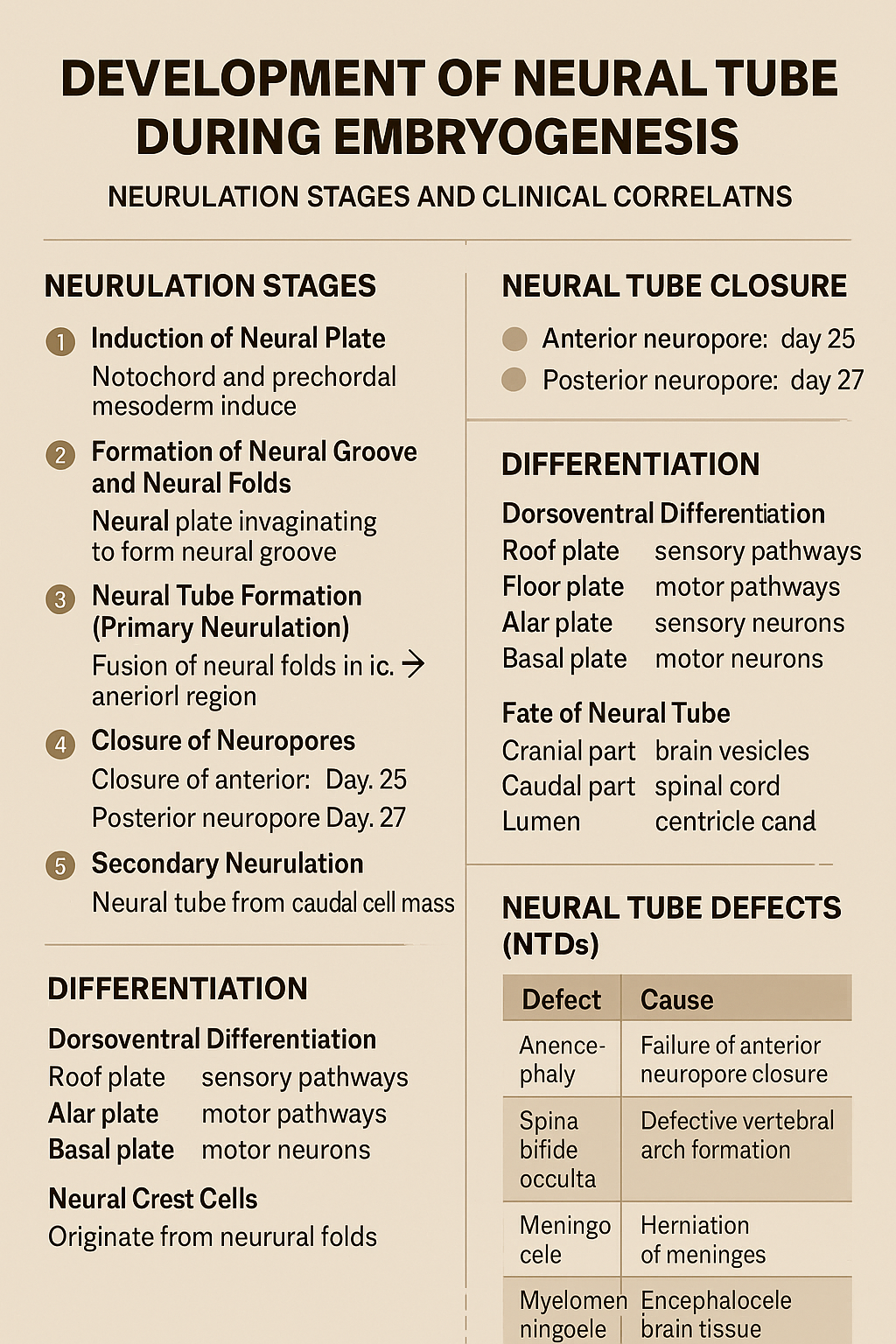
* **Day 25**: Closure of anterior (cranial) neuropore
* **Day 27**: Closure of posterior (caudal) neuropore
---
## **Phases of Neural Tube Development**
### **1. Induction of Neural Plate**
* The **notochord** and **prechordal mesoderm** induce overlying **ectoderm**.
* Ectoderm thickens to form the **neural plate**.
**Key signaling molecules**:
* Sonic hedgehog (Shh)
* Noggin
* Chordin
* Follistatin
---
### **2. Formation of Neural Groove and Neural Folds**
* Central neural plate invaginates → **neural groove**.
* Lateral edges elevate → **neural folds**.
* Neural folds approach each other dorsally.
---
### **3. Neural Tube Formation (Primary Neurulation)**
* Neural folds fuse in the **midline**, starting in the **cervical region**.
* Fusion proceeds cranially and caudally.
* Temporary openings remain:
* **Anterior (cranial) neuropore**
* **Posterior (caudal) neuropore**
---
### **4. Closure of Neuropores**
* **Anterior neuropore closes (Day 25)** → brain development
* **Posterior neuropore closes (Day 27)** → spinal cord development
Failure of closure leads to **neural tube defects**.
---
### **5. Secondary Neurulation**
* Occurs in the **caudal region (below S2)**.
* Neural tube forms from **caudal cell mass**.
* Important for formation of:
* Conus medullaris
* Filum terminale
---
## **Differentiation of the Neural Tube**
### **Neural Tube Wall Layers**
1. **Ventricular (ependymal) layer** – neuroepithelial cells
2. **Mantle layer** – gray matter
3. **Marginal layer** – white matter
---
### **Dorsoventral Differentiation**
* **Roof plate** → sensory pathways
* **Floor plate** → motor pathways
* **Alar plate** → sensory neurons
* **Basal plate** → motor neurons
* **Sulcus limitans** separates alar and basal plates
---
## **Fate of Neural Tube**
* **Cranial part** → brain vesicles
* Prosencephalon
* Mesencephalon
* Rhombencephalon
* **Caudal part** → spinal cord
* **Lumen** → ventricles and central canal
---
## **Neural Crest Cells (Related Event)**
* Originate from neural folds
* Migrate extensively
* Form:
* Peripheral nervous system
* Melanocytes
* Adrenal medulla
* Craniofacial cartilage
---
## **Clinical Correlation: Neural Tube Defects (NTDs)**
| Defect | Cause |
| -------------------- | ------------------------------------- |
| Anencephaly | Failure of anterior neuropore closure |
| Spina bifida occulta | Defective vertebral arch formation |
| Meningocele | Herniation of meninges |
| Myelomeningocele | Herniation of spinal cord + meninges |
| Encephalocele | Herniation of brain tissue |
**Risk factors**:
* Folic acid deficiency
* Maternal diabetes
* Valproate exposure
**Prevention**:
* Folic acid 400–800 µg/day (preconception)
---
### **One-Line Exam Summary**
> The neural tube forms from ectoderm by neurulation during weeks 3–4 and develops into the brain and spinal cord.
If you want, I can also provide **exam-oriented MCQs, flowcharts, or comparison tables** for neurulation.